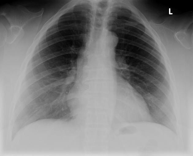
Radiology – Case 3
An X-ray was taken in a 30 year old male who presented with a history of left sided chest pain. What is the finding is the X-ray given above? What is the condition that caused the above finding. What are the clinical features?
Note: The finding may not be related to the presenting complaint.
Image Credits : Radiopedia
13 Comments


i think its a case of mitral stenosis
aortic knob
bilateral absence of clavicle,lvh,prominent aortic knuckle…cleidocranial dystosis…
this skiagram is taken during expiration .causes of left sided chest pain could be multiple.
to me it is looking lyk a collapse middle lobe silhoutte sign positive it could also be a hilar lymphadenopathy
ALSO CLAVICLES ARE ABSENT
two things observed
1] ground glass opacity over both lower hilum s/o consolidation?
2] either fundal gas shadow or query air under left diaphram s/o ?? peptic perforation
Descending aortic aneurysm
fract Left clavicle
prominent aortic knuckle, right ventricle bulge, enlarged heart, consolidation in lower lobe of lung, right side clavicle is not seen..faint shadow of left clavicle is seen..
hello i say this is a/p veiw dan nor p/a view,i can make out some thing in d postier mediastinal shodow probebly behibd heart probeblr acase f diaphgramatic hernia wr bowel loop in mediasinum
seems to be consolidation of both lower lobe..
aortic knuckle s/o aortic aneurism,b/l lower lobe hazziness s/o pneumonia/pulmonary oedema,cardiomegaly,widened mediastinal shadow,absent clavicle
b/l ll consolidation wid increased hillar vessel markings probably pulmonary ht …