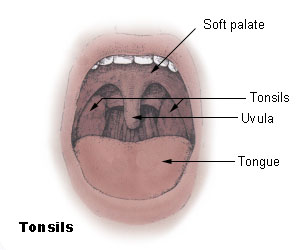
Anatomy of palatine tonsil
- The palatine tonsil is an ovoid mass of lymphoid tissue located in the oropharynx between the anterior and posterior pillars
- It has a 2 surfaces – medial and lateral and 2 poles – upper and lower
Medial surface
- It is lined by stratified squamous non keratinising epithelium which dips into the crypts
- The crypts are 12-15 in number
- Secondary crypts arise from the primary crypts and extend into the substance of the tonsil
- On of the crypts located in the upper part are larger than the rest – crypta magna
- It represents the ventral part of second pharyngeal pouch
- The crypts serve to increase the surface area of the tonsil
- The crypts may be filled witth cheesy material – epithelial debris, food particles and bacteria
Lateral surface
- It is covered by the fibrous capsule of the tonsil
- The tonsillar bed is separated from the capsule by loose areolar tissue
- This makes it is easy to dissect the tonsil from its bed during tonsillectomy
- It is the site of collection of pus in peritonsillar abscess (quinsy)
- Some fibers of palatoglossus and palatopharyngeus gets attached to capsule of tonsil
Upper pole
- It extends into the soft palate
- There is a semilunar fold of mucous membrane which covers the medial part of the upper pole
- It extends from anterior pillar to posterior pillar
- It encloses a potential space – supratonsillar fossa
Lower pole
- It is attached to the tongue
- A triangular fold of mucous membrane extends from the anterior tonsillar pillar to the lower pole
- It encloses a space – anterior tonsillar space
- The lower pole is separated from the tongue by the tonsillolingual sulcus
- This sulcus may harbour carcinoma
Bed of tonsil
- It is formed by the 2 muscles
- Superior constrictor
- Styloglossus
- View Main article – Structures related to bed of tonsil
Blood supply
- The tonsil is supplied by branches of external carotid artery
- See main article – Blood supply of tonsil
Venous drainage
- Blood from the tonsil drains into the paratonsillar vein which in turn drains into the common facial vein and pharyngeal venous plexus
Lymphatic drainage
- Lymphatics from the tonsil pierce the superior constrictor and drain into the upper cervical lymph nodes especially jugulodigastric (tonsillar) lymph node
- Enlarged non tender jugulodigastric lymph node is a sign of chronic tonsillitis
Nerve supply
- Lesser palatine branch of sphenopalatine ganglion
- Glossopharyngeal nerve
Functions of tonsil
- It has a protective function in that it prevents entry of pathogens through the nasal and oral route
- The crypts on the surface of the tonsil serve to increase the surface area and increase the efficiency of protection against pathogens
- It forms a part of Waldeyer’s lymphatic ring


