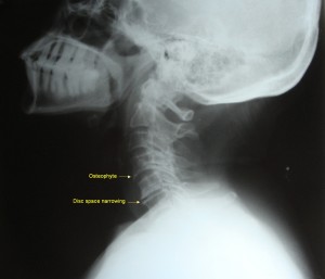
Spondylotic changes – cervical vertebrae – X-ray

X-ray neck – lateral view – showing spondylotic changes
Click on image for an enlarged view
Spondylotic changes to look for in lateral view radiograph of neck:
- Osteophytes
- Disc space narrowing
- Loss of cervical lordosis
- Uncovertebral joint hypertrophy
- Apophyseal joint osteoarthritis
- Decreased vertebral canal diameter
Other imaging modalities for diagnosing cervical spondylosis:
- CT scan
- CT myelography
- MRI – investigation of choice


