
60 year old man with acute pancreatitis- Medicine MCQ
A 60 year old man presented to the OPD with symptoms suggestive of acute pancreatitis. He consumes high quantities of alcohol regularly. His symptoms started 4 days ago. But he continued to consume alcohol. He was admitted for further evaluation. Presently, he has severe vomiting. He also complains of dizziness when standing. Examination revealed tenderness in the epigastrium and right hypochondrium. A reddish discolouration is noted in the flanks. Which of the following statements regarding the patient is most accurate?
A. The patient should be evaluated for concomitant appendicitis
B. USG is likely to demonstrate pseudocyst of pancreas
C. Contrast CT scan of the abdomen will reveal severe necrotising pancreatitis
D. Pancreatic calcification will be seen in X-ray abdomen
Correct answer : C. Contrast CT scan of the abdomen will reveal severe necrotising pancreatitis
The history and clinical features (Abdominal tenderness, hypotension due to blood loss, Grey Turner’s sign) are suggestive of acute pancreatitis with pancreatic necrosis. Hence a contrast CT scan of the abdomen will reveal severe necrotising pancreatitis.
Eponymous clinical signs in acute pancreatitis
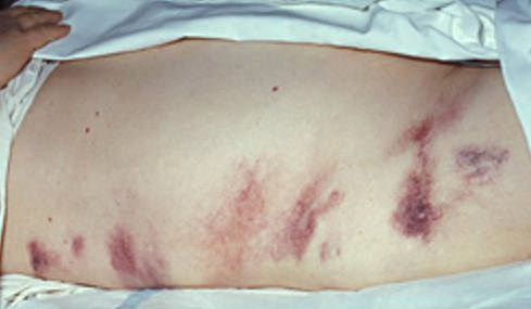 Grey Turner’s sign
- Reddish / bluish discolouration of the flanks is known as Grey Turner’s sign.
- It is named after George Grey Turner, a British surgeon.
- It is seen in cases of acute pancreatitis with pancreatic necrosis and intra abdominal bleeding.
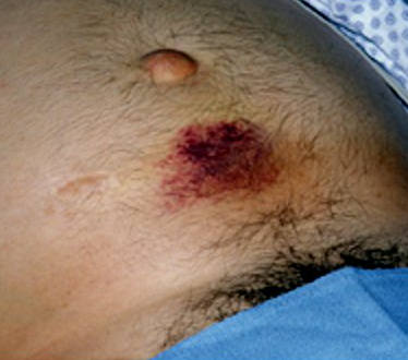 Cullen’s sign (in this image, the discolouration is seen below the umbilicus instead of around it)
- Another feature of pancreatic necrosis is a bluish discolouration around the umbilicus, known as Cullen’s sign.
- It is named after Thomas Stephen Cullen, a Canadian gynecologist who first described the sign in a case of ruptured ectopic pregnancy in 1916.
- Both these signs take 1-2 days to develop in a case of acute pancreatitis.
CT findings in acute pancreatitis
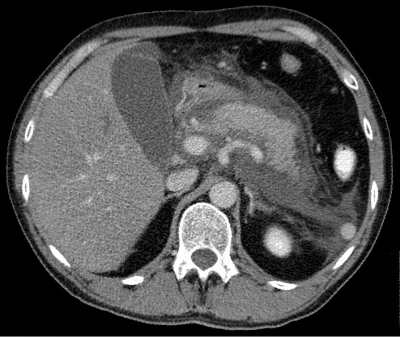 Acute pancreatitis – CT scan
- Focal / diffuse enlargement of pancreatic parenchyma
- Indistinct margins
- Density changes due to presence of oedema
- Retroperitoneal stranding
Elimination of the other options
- Pseudocyst of pancreas usually develops a few weeks after the onset of pancreatitis
- The features are not suggestive of appendicitis
- Pancreatic calcification is seen in chronic pancreatitis
Image credits:
- Grey Turner’s sign – Herbert L. Fred, MD and Hendrik A. van Dijk via http://cnx.org/content/m14942/latest/
- Cullen’s sign – Herbert L. Fred, MD and Hendrik A. van Dijk via http://cnx.org/content/m14904/latest/
- Acute pancreatitis – CT scan – Hellerhoff via http://en.wikipedia.org/wiki/File:Akute_exsudative_Pankreatitis_-_CT_axial.jpg


So helpfull