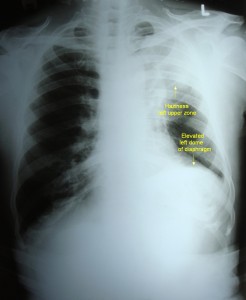
Mass lesion – Left lungs – X-ray

Mass lesion – left lung
Click on image for an enlarged view
- X-ray chest posteroanterior view showing homogenous haziness in left lung – upper and mid zones
- Elevation of left dome of diaphragm – due to volume loss secondary to bronchus obstruction / diaphragmatic palsy secondary to phrenic nerve involvement
- Mediastinal shift to left side
- Hyperinflated right lung
Impression – Bronchogenic carcinoma
Factors in favour of diagnosis of bronchogenic carcinoma
- Homogenous haziness (Tuberculosis can cause haziness of upper zone – due to fibrosis, but the appearance is non homogenous)
- Volume loss can occur due to compression of bronchus by the tumour
- Mediastinal shift due to volume loss
- Phrenic nerve infiltration can cause diaphragmatic palsy


